Differential Diagnosis in Neuroimaging: Brain and Meninges
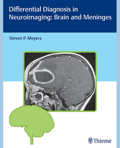
Differential Diagnosis in Neuroimaging: Brain and Meninges
Summary
The distinctive 'three-column table plus images' format is easy to incorporate into clinical practice, setting this book apart from larger, disease-oriented radiologic tomes. The layout enables readers to quickly recognize and compare abnormalities based on high-resolution images.
This visually rich resource is a must-have diagnostic tool for radiologists, neurosurgeons, and neurologists, and residents and fellows. The highly practical format makes it ideal for daily rounds, as well as a robust study guide for physicians preparing for board exams.
On CT, the appearance of brain tissue depends on the mAs and kVp used. Immature myelin in neonates and infants has lower attenuation than myelin in older children. In adults, the cerebral cortex has an intermediate attenuation that is slightly higher relative to normal white matter. The imaging changes seen with myelin maturation are more optimally seen with MRI than with CT.
The MRI features ofsubdural hematomasare variable, although the appearances can progress in patterns similar to those for intraparenchymal hematomas. Chronic subdural hematomas often have a low-intermediate signal on T1-weighted images and a high signal on T2-weighted images.Subarachnoid hemorrhageis often difficult to see on T1- and T2-weighted images, although it can be sometimes identified on long TR/short TE (proton density-weighted images) or FLAIR images.