Imaging of Cerebrovascular Disease: A Practical Guide
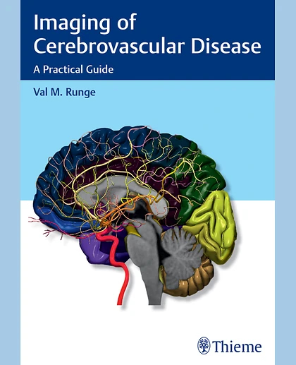
Imaging of Cerebrovascular Disease: A Practical Guide
Summary
Imaging plays an integral role in the diagnosis and intervention of debilitating, often fatal vascular diseases of the brain, such as ischemic and hemorrhagic stroke, aneurysms, and arteriovenous malformations (AVMs). Written by a world renowned neuro-radiologist and pioneer in the early adoption of magnetic resonance (MR) technology,Imaging of Cerebrovascular Diseaseis a concise yet remarkably thorough textbook that advances the reader's expertise on this subject.
Key Features:In-depth discussion of medical and neuroradiologic/neurosurgical interventions, focusing on the use of imaging prior to, during, and following treatmentComprehensive text enhanced with more than 700 high-quality imagesDetailed evaluation of normal brain anatomy, as well as gyral anatomy in brain ischemia, an important subtopicAdvantages, disadvantages, mortality, and morbidity of surgery (clipping) versus endovascular techniques (coiling and flow diversion) for aneurysms
Presented in a style that facilitates cover-to-cover reading, this is an essential tool for residents and fellows, and provides a robust study guide prior to sitting for relevant certification exams. It is also a quick, invaluable reference for radiologists, neurosurgeons, and neurologists in the midst of a busy clinical day.
Chapter 1 Technologic Innovations in MR and CTChapter 2 Normal AnatomyChapter 3 HemorrhageChapter 4 IschemiaChapter 5 AneurysmsChapter 6 Vascular Malformations and Other Vascular Lesions