Differential Diagnosis in Neuroimaging: Head and Neck
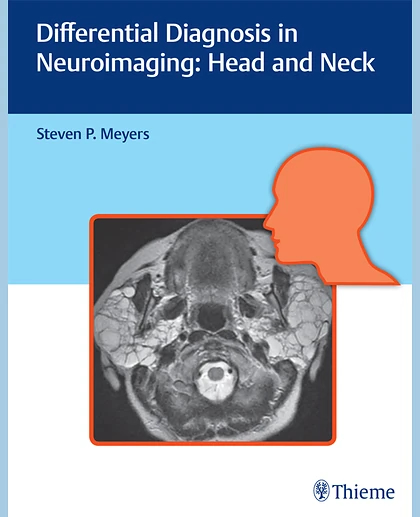
Differential Diagnosis in Neuroimaging: Head and Neck
Summary
The distinctive 'three-column table plus images' format is easy to incorporate into clinical practice, setting this book apart from larger, disease-oriented radiologic tomes. This layout enables readers to quickly recognize and compare abnormalities based on more than 1,500 high-resolution images. Chapters cover skull imaging, temporal bone imaging, orbital imaging, paranasal imaging, suprahyoid neck imaging, and infrahyoid neck imaging, for a full spectrum of head and neck pathologies.
This visually rich resource is a must-have diagnostic tool for residents, fellows, and practitioners in radiology, otolaryngology-head and neck surgery, and neurosurgery. The highly practical format makes it ideal for daily rounds, as well as a robust study guide for physicians preparing for board exams.
1 Skull and Temporal Bone2 Orbit3 Paranasal Sinuses and Nasal Cavity4 Suprahyoid Neck5 Infrahyoid Neck6 Lesions That Can Involve Both Suprahyoid and Infrahyoid Neck7 Brachial Plexus
Table 2.1Congenital and developmental abnormalities